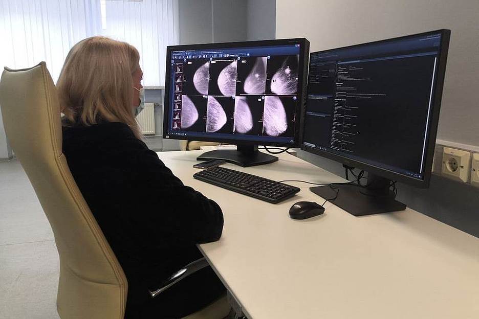
64% of radiologists agreed with the results of the algorithms of Yekaterina Alexandrov | 09.11.2020

Over the past six months increased the number of radiologists, who are active users of medical services based on computer vision. The number of skeptical doctors to
artificial intelligence technology has dropped to 36%. These results are reported by Sergey Morozov, the chief specialist of the Moscow radiation and instrumental diagnostics, Director of the Center for diagnosis and
telemedicine.
The results of the work of AI services in the framework of the Moscow experiment on introducing computer vision in x-ray diagnostics to assess the five hundred radiologists. Moscow experiment on introduction
computer vision in x-ray diagnostics conducted from February 2020. This is a large-scale scientific study of the applicability and quality of artificial intelligence to solve practical problems
health. The results of the experiment will be announced next year.
New data showed that 64% of the radiologists agree with the results of the algorithms of artificial intelligence. The rest of the users still dissatisfied with the quality of medical treatment
images — the results, according to them, incorrect. Some doctors believe that the occurrence of false-positive and false-negative results in the processing and analysis of medical images
artificial intelligence is associated with the quality of IT development, and it needs “dobuchin” algorithms. The real causes of misdiagnosis Sergey Morozov calls the problem
developer: gaps in the quality management system and methodically incorrect matching of datasets for learning algorithms.
“Not all experts are quick to support new technologies, every IT product adapts to changes across the curve of innovation. In addition, there are doctors who Express concerns
the fact that the profession of a radiologist in the future under the threat. This is not true. It is obvious that in the future radiologists will work with a lot of difficult research,
as growing volumes of diagnostic and research methods. While the share of artificial intelligence will be routine, monotonous tasks, such as preventive research,” — says Sergey
Frost.
The high flow studies, resulting in staff shortages will be able to solve the computer vision technology, aimed at reducing the time of the description of medical images. In
currently 33% of radiologists believe that algorithms will be able to reduce time to prepare reports. However, the other 32% of radiologists believe in the opposite: in their view, the time to
description increased due to the AI, as not all functionality of the services modified. However, 46% of doctors believe that AI services will help to reduce the omissions of clinically significant
differences. In the future, the operator of the experiment — Center of diagnostics and telemedicine — is considering the creation of personalized worklists for doctors depending on their specialty, within
which radiologists will be able to choose certain algorithms, but this model needs to be tested.
According to the research of the Centre for diagnostics and telemedicine, the average processing time for medical imaging services is 10 minutes. Standards of health for the planning description
x-ray studies is allocated 24 hours. “Real time training may take less time due to remote research papers, a concentration of expertise in
Reference centre and automation of the process,” said Anton Vladzimirsky, Deputy Director on scientific work of the Centre for diagnostics and telemedicine. This is especially true for situations when
the duration of the training x-ray may achieve a week, and records for research are opened, pending the presence of a doctor on the spot.
Another objective of the digital project, as was concluded Anton Vladzimirsky, is to reduce the burden on doctors for the treatment of large flow routine studies with a low percentage of detection of important
pathologies. According to him, three thousand viewed fluorogram it is possible to identify only one case with signs of tuberculosis. For this reason, in the experiment included such routine and mass
investigations as fluorography, mammography, lung cancer screening, which can be automated using computer vision techniques. Along with this, in the future, some functions
radiologists can be transferred to other professionals. In particular, the lungs x-ray with automated markup of pneumothorax’ll be able to do the doctor-resuscitator,
which, in accordance with the professional standard, has the right to the diagnosis.
Artificial intelligence, medicine


